
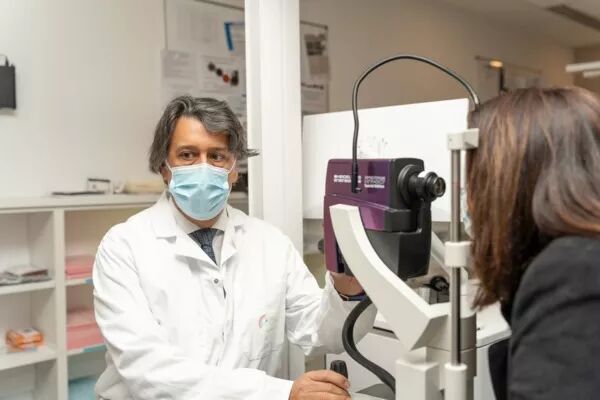
Pr Eric Souied 🇬🇧
Techniques and Examinations
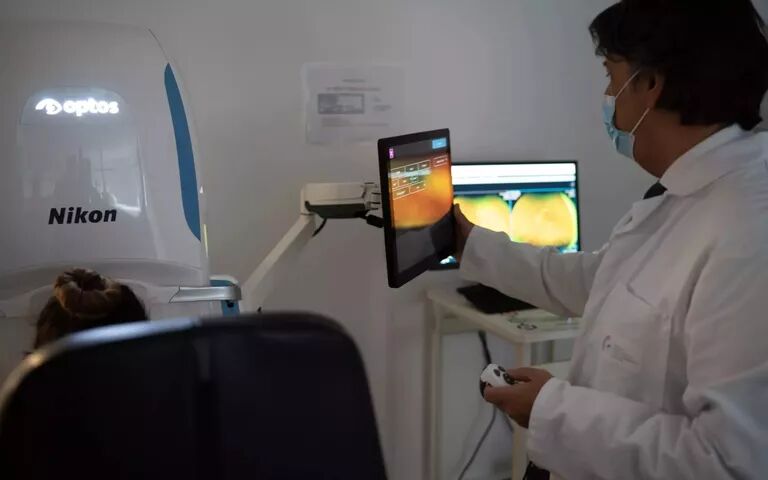
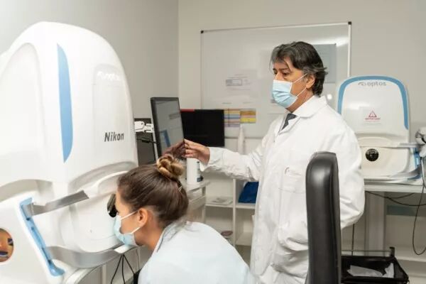
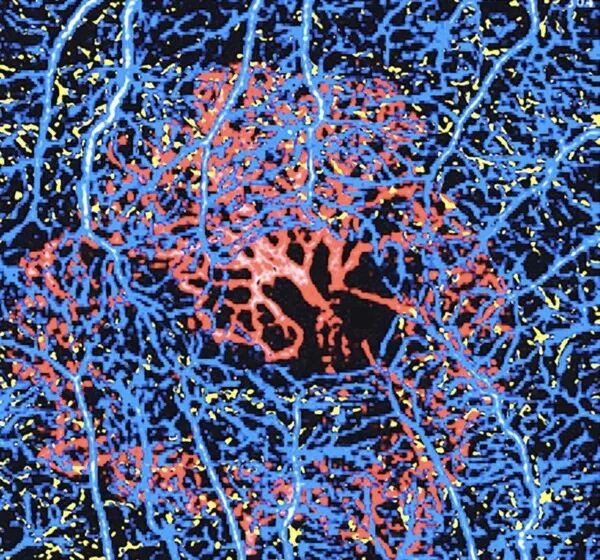
Angiography

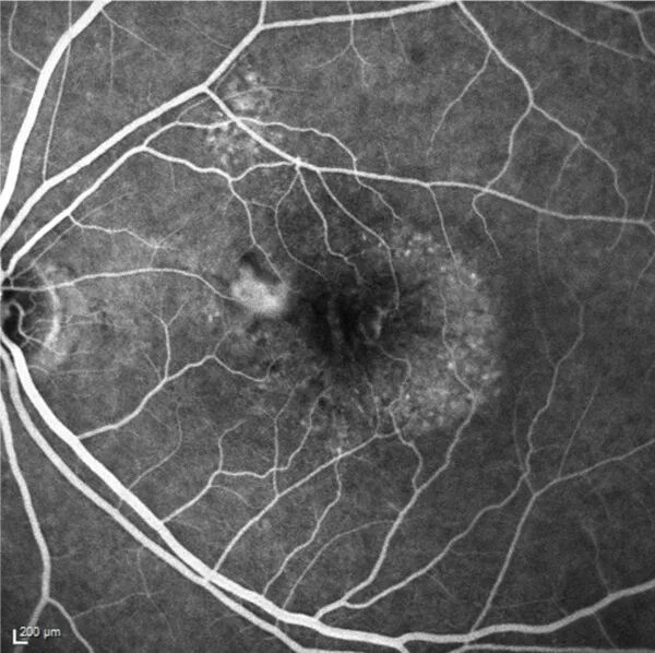

Angiography
Angiography is a retinal imaging examination that allows the study of blood vessels not visible on standard fundus photographs. Its purpose is to detect vascular disorders and assess the quality of retinal vessels. It remains the gold standard for diagnosing most retinal and macular pathologies.
Angiography is a safe examination with very few risks of adverse effects.
There are two types of angiography: fluorescein angiography and indocyanine green (ICG) angiography, which use different dyes and provide different views of the retina and choroid.
Ultra-widefield imaging

Ultra-widefield imaging
Ultra-widefield imaging (UWF) engineering provides the capability to capture nearly the entire fundus in a single image.
This allows for the visualization of all lesions on 200-degree images without the need for assembling multiple 50-degree images, which optimizes diagnosis and makes it easier to detect vascular abnormalities in the retinal periphery
OCT
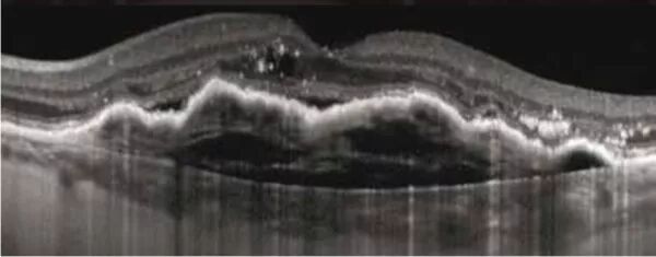

OCT
Optical Coherence Tomography (usually referred to by its acronym OCT) is a medical imaging technique that provides cross-sectional images at various depths and reconstructs the entire eye using a principle similar to that of a scanner. It uses non-ionizing infrared radiation and poses no risk.
It can provide images of the retina, optic nerve, cornea, anterior chamber, and iridocorneal angle. OCT is used for the detection of glaucoma, age-related macular degeneration (AMD), and diabetic macular edema.
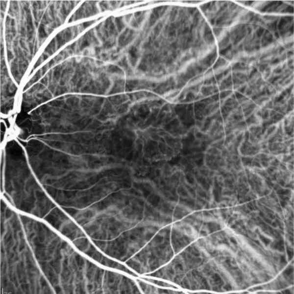
OCT - Angiography
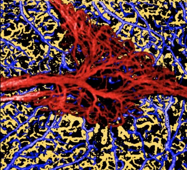
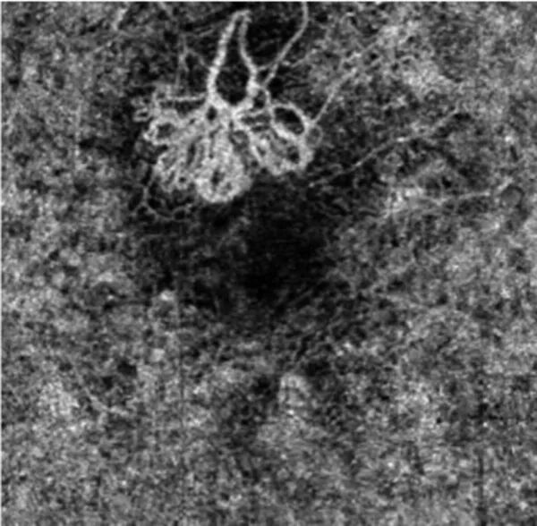
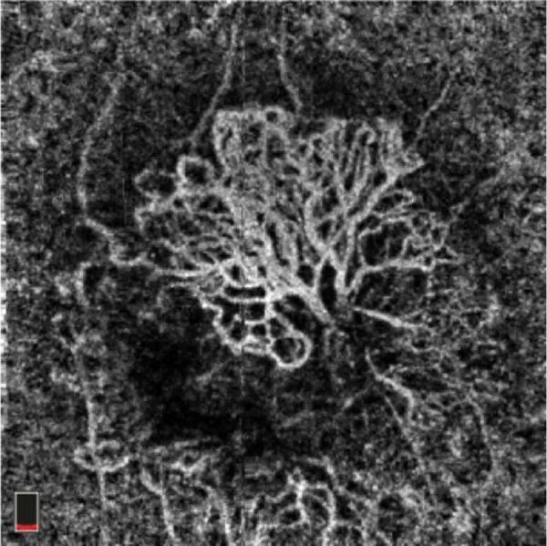
OCT - Angiography
OCT-Angiography, or OCTA, is an angiography technique combined with optical coherence tomography (OCT), and it does not require any contrast agent injection. This relatively recent technology has enabled the discovery of new pathologies and the early detection of subclinical macular disorders